


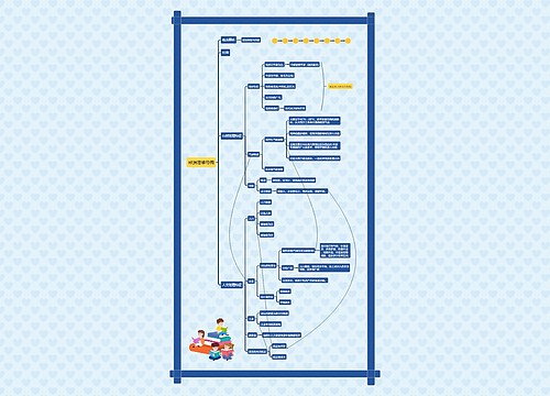
digestive system思维导图

消化系统详解
树图思维导图提供 digestive system 在线思维导图免费制作,点击“编辑”按钮,可对 digestive system 进行在线思维导图编辑,本思维导图属于思维导图模板主题,文件编号是:a6d9eb7f9ffe1698e9488f60a0033e3a
思维导图大纲
digestive system思维导图模板大纲
Small intestin
Duodenum
Upper part: the beginning is called duodenal bulb, which is the most common site of duodenal ulcer and its perforation.
Descending part
Horizontal part
Ascending part
Jejunum and ileum
Jejunum is often located in the left lumbar region and umbilical region, and ileum is often located in the umbilical region, right inguinal region and pelvis.
Oral cavity
Lip
Cheek
There is a parotid duct nipple on the buccal mucosa opposite to the crown of the maxillary second molar.
Palate
Hard hire: the first 2/3 of the palate.
Soft hire: located in the back 1/3 of the palate.
Teeth
Types of teeth
Deciduous tooth
Permanent teeth
The shape of the tooth
Crown, root, neck
Dental tissue
Dentin, enamel, cementum, dental pulp
Periodontal tissue
Periodontal ligament, alveolar bone, gingiva
Tongue
The shape of the tongue
Divided into the root of the tongue and the body of the tongue
Tongue mucosa
Lingual papilla
Filamentous nipple
Fungal nipple
Phylloid papilla
Contour nipple
Sublingual caruncle, frenulum, sublingual fold, sublingual gland, etc.
Lingual muscle
Medial lingual muscle
Longitudinal muscle, transverse muscle, vertical muscle
Extralingual muscle
Genioglossus muscle, hyoid muscle, stylohyoid muscle
Salivary gland
Large salivary gland
Parotid gland, submandibular gland, sublingual gland
Minor salivary gland
Labial gland, buccal gland, palatal gland, tongue gland
Pharynx
Nasopharynx
Oropharynx
Laryngopharynx
large intestine
Cecum
The beginning of the large intestine, about 6~8cm, its lower end is the blind end, the ascending colon continues, and the left side is connected with the ileum. The cecum is located in the right iliac fossa and its body surface is projected above the lateral half of the inguinal ligament.
Appendix
The body surface projection point of the root of the appendix: usually at the intersection of 1/3 of the line between the right anterior superior iliac spine and the umbilical cord, this point is called McBurney point. Sometimes it is also expressed as the Lanz point, that is, the intersection of the right and middle 1/3 of the line connecting the left and right anterior superior iliac spine.
colon
Ascending colon
Transverse colon
Descending colon
Sigmoid colon
Rectum
Anal canal
Main function: absorb water, vitamins and inorganic salts, and form food residues into feces and discharge them out of the body.
esophagus
Division
Neck
About 5cm, the segment from the beginning of the esophagus to the plane of the sternal jugular notch.
Chest
About 18~20cm, located between the plane of the sternal jugular notch and the esophageal hiatus of the diaphragm.
Abdomen
1~2cm, from esophageal hiatus to cardia.
Constriction
The first stenosis
The beginning of the esophagus is equal to the level of the lower margin of the sixth cervical vertebra, about 15cm from the central incisor.
The second stenosis
The esophagus is at the intersection of the left main bronchus, equivalent to the level between the 4th and 5th thoracic vertebrae, and about 25cm from the central incisor.
The third stenosis
The esophagus passes through the esophageal hiatus of the diaphragm, equivalent to the level of the 10th thoracic vertebra, about 40cm from the central incisor.
Stomach
Division
Cardiac part
The cardia is located on the left side of the 11th thoracic vertebra.
Gastric fundus (gastric fornix)
Gastric body
Pyloric part (gastric antrum)
The pylorus is located on the right side of the first lumbar vertebra.
Position
When the stomach is moderately full, most of it is located in the left costal region and a small part in the epigastric region.
Adjacent: the right part of the anterior wall of the stomach is adjacent to the left lobe and square lobe of the liver, and the left part is adjacent to the diaphragm, which is covered by the left costal arch. Below the xiphoid process, part of the anterior wall of the stomach is directly attached to the ventral anterior wall. The posterior wall of the stomach is adjacent to the pancreas, transverse colon, upper left kidney and left adrenal gland, and the fundus of stomach is adjacent to the diaphragm and spleen.
Function
Receive food, secrete gastric juice, endocrine function
Liver
Position
Most of them are located in the right costal region and epigastric region, and a small part in the left costal region.
Form
Diaphragmatic surface
There are falcate ligament and coronal ligament on the diaphragmatic surface of the liver.
The falciform ligament is in a sagittal position, dividing the liver into left and right lobes.
The part between the two layers of the coronal ligament of the posterior part of the diaphragm without peritoneal cover is called the bare area.
The visceral surface of the liver
Three H-shaped trenches
Left longitudinal sulcus
Anterior: fissure of round ligament of liver, with traces of round ligament of liver and umbilical vein.
Posterior: venous ligament fissure, traces of venous ligament and venous catheter.
Transverse sulcus (hilum of liver)
There are left and right hepatic ducts, left and right branches of proper hepatic artery, left and right branches of hepatic portal vein and nerves and lymphatic vessels, also known as the first hepatic hilum.
These structures entering and leaving the hilum of the liver are surrounded by connective tissue to form the hepatic pedicle.
Right longitudinal sulcus
Anterior: gallbladder fossa.
Posterior: vena cava sulcus (second hepatic hilum), where the left, middle and right hepatic veins flow out of the liver into the inferior vena cava.
Extrahepatic biliary system
Bile duct
Common bile duct
Common hepatic duct
Cystic duct
Common hepatic duct
Left hepatic duct
Right hepatic duct
Gallbladder triangle
A triangular area surrounded by the cystic duct, the common hepatic duct, and the visceral surface of the liver.
Gallbladder
Division
Gallbladder floor
Gallbladder body
Neck of gallbladder
Cystic duct
Function
Store, concentrate bile
Function
Participate in the synthesis, transformation and decomposition of proteins, lipids, sugars and vitamins.
Participate in the transformation and detoxification of hormones, drugs and other substances.
Important functions such as bile secretion, phagocytosis, defense and hematopoiesis during embryonic period
Pancreas
Composition
Exocrine part (glandular cell)
Can secrete pancreatic juice and contain a variety of digestive enzymes (such as protease, lipase and amylase). It can decompose and digest proteins, fats and sugars.
Endocrine part (islet)
Mainly secrete insulin and regulate the concentration of blood glucose.
Position
Located in the epigastric region and the left costal region, transversely placed in front of the 1st ~ 2nd lumbar vertebrae, and close to the posterior abdominal wall.
Division
Head of pancreas
Neck of pancreas
Body of pancreas
Tail of pancreas
Pancreatic duct
Pancreatic duct
Merge with the common bile duct to form the ampulla of hepatopancreas, which opens in the duodenum with large breast and head
Accessory pancreatic duct
It opens in the small papilla of the duodenum and mainly drains pancreatic juice from the anterior upper part of the head of the pancreas.
Composition
Digestive canal
Upper digestive tract: from oral cavity to duodenum.
Lower digestive tract: below jejunum.
Digestive gland
Large digestive glands: large salivary glands, liver and pancreas.
Small digestive gland: labial gland, buccal gland, tongue gland, esophageal gland, gastric gland, intestinal gland, etc.
Contains taste buds, which are taste receptors思维导图模板大纲
Tasteless bud, tasteless function思维导图模板大纲
Stricture is a common site for retention of esophageal foreign bodies and esophageal cancer.思维导图模板大纲
相关思维导图模板


树图思维导图提供 A Secure System For Pervasive Social Network-Based Healthcare 在线思维导图免费制作,点击“编辑”按钮,可对 A Secure System For Pervasive Social Network-Based Healthcare 进行在线思维导图编辑,本思维导图属于思维导图模板主题,文件编号是:3fe009af1ca6673fe665df5e689aa6ad

树图思维导图提供 社会主义基本经济制度The basic socialist economic system 在线思维导图免费制作,点击“编辑”按钮,可对 社会主义基本经济制度The basic socialist economic system 进行在线思维导图编辑,本思维导图属于思维导图模板主题,文件编号是:e73ec0d3f86f78a33e45762aea9cae5f


 上海工商
上海工商